Dental technology
3D Imaging, Digital X-Rays & Digital Impressions
We’re adding 3D imaging, digital X-rays and digital impressions, for more comfortable visits and fewer appointments. Here’s what each one does, how dentists keep radiation low, and how it helps with implants, root canals, braces and crowns.
- 3D imaging being added
- Digital X-rays
- Digital impressions
- Caring for Murrieta since 1991
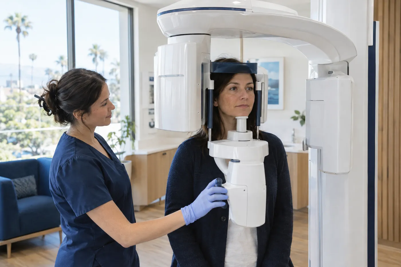
What is 3D dental imaging?
3D dental imaging, usually done with cone beam CT (CBCT), is a type of X-ray that rotates around your head and builds a three-dimensional view of your teeth, jaws, nerves and sinuses. Dentists use it when standard two-dimensional X-rays can’t provide the information they need, such as when planning dental implants.
What technology is new at our Murrieta office?
We’re adding three tools: 3D imaging, digital X-rays and digital impressions. As our letter to patients explains, the goal is more comfortable visits and fewer appointments.
Each one does a different job. Digital X-rays are the everyday pictures of your teeth. 3D imaging is a deeper look saved for specific questions. Digital impressions replace trays of impression material with a small scanner for many kinds of treatment.
Your records stay right here, and new images join the digital records our office already keeps. Read more about our office and technology.
What is a cone beam CT image?
A cone beam CT (CBCT) image is a 3D X-ray of your teeth, jaws and the structures around them. The FDA explains that the machine rotates around you, capturing data with a cone-shaped X-ray beam, and creates three-dimensional information instead of the flat, two-dimensional view of a standard dental X-ray.
That 3D view lets your dentist see the jawbone, nerves, sinuses and tooth roots from every angle, which a flat image can’t fully show. Cleveland Clinic notes CBCT is often used for implant planning.
According to the FDA, dentists also use CBCT to look at abnormal teeth, evaluate the jaws and face, and help diagnose cavities, root canal problems and dental injuries. For the scan, you stay still while the machine moves around your head.
Injured? Emergency care comes first
For a serious injury to the face, facial swelling that affects your breathing, swallowing or vision, a high fever with swelling, or bleeding that won’t stop, call 911 or go to the nearest ER.
At a glance
Three technologies at a glance
How digital X-rays, 3D imaging and digital impressions differ.
| Digital X-rays | 3D imaging (CBCT) | Digital impressions | |
|---|---|---|---|
| What it is | Flat images captured by an electronic sensor | A 3D X-ray scan of the teeth, jaws, nerves and sinuses | A 3D model of your teeth made with a handheld scanner |
| Common uses | Checkups, cavities between teeth, roots and bone | Implant planning, abnormal teeth, jaw evaluation, some root canal diagnosis | Crowns, bridges, aligners, bite guards and implant guides |
| Radiation | Very low; less than film | More than standard dental X-rays, less than other CT exams | None; it’s an optical scan, not an X-ray |
| How often | Based on your exam, history and risk | Only when standard X-rays can’t answer the question | When a treatment needs a model of your teeth |
How are digital X-rays different from film?
Digital X-rays use an electronic sensor in place of film, and the image appears on a computer screen. Cleveland Clinic notes that digital X-rays use much less radiation than traditional film X-rays, and the ADA lists digital sensors among the ways dentists keep exposure low.
They’re still the everyday tool for checkups. Bitewing X-rays show cavities between teeth and below the gumline, and periapical X-rays show the roots and surrounding bone, according to Cleveland Clinic. Because the picture is on a screen, your dentist can enlarge it and walk you through what they see.
How often you need X-rays depends on you. The ADA’s MouthHealthy site says the schedule depends on your oral health, age, disease risk and any symptoms, and that X-rays should be taken only when your dentist believes they will provide needed information.

What are digital impressions?
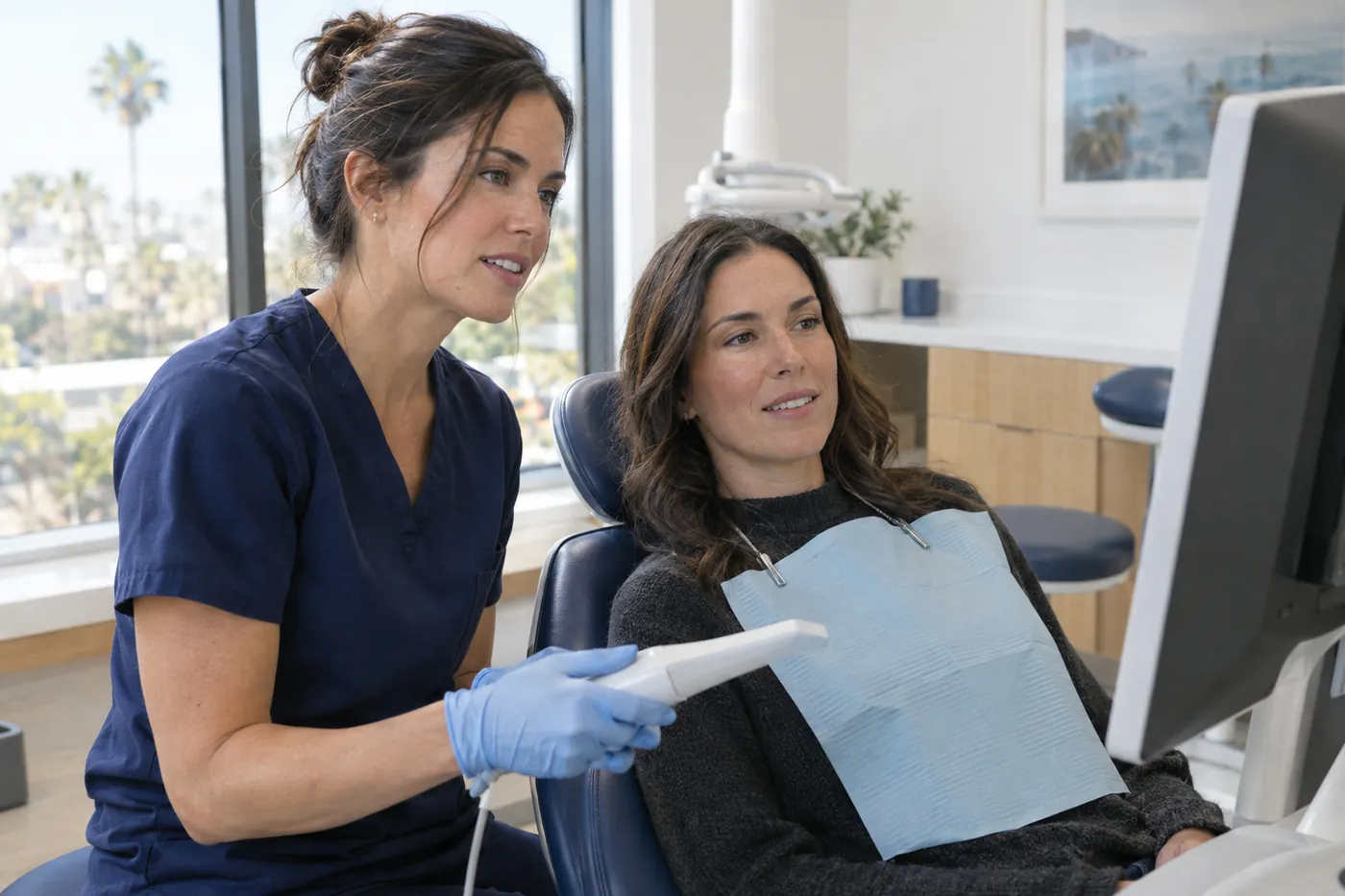
A digital impression is a 3D model of your teeth made with a small handheld scanner instead of a tray of impression material. Your dentist moves the scanner around your teeth, and the model builds on a screen.
Traditional impressions mean biting into a tray of soft material and holding still while it sets, which some people find uncomfortable or hard on their gag reflex. The ADA notes that scanners may provide a faster and more comfortable experience than the traditional method. A 2024 study of children and teens also found more comfort and less gagging with scans than with conventional impressions.
According to the ADA, dentists use digital impressions to create crowns, inlays, onlays, bridges, clear aligners, bite guards and implant surgical guides. A scan captures what researchers call an optical impression, made with light, so it isn’t an X-ray.
Is dental imaging safe? How radiation is kept low
Dental X-rays use very low doses of radiation, and the ADA’s MouthHealthy site says the risk of harmful effects is very small. 3D imaging is different: the FDA notes that dental CBCT typically delivers more radiation than standard dental X-rays, though generally less than other CT exams. That’s why CBCT is used only when it’s needed.
Here are the principles behind how dentists decide on imaging, from FDA and ADA guidance:
- Only when it will change your care. The ADA and FDA’s patient-selection guidance says X-rays should be taken when the information is expected to affect your treatment, decided after an exam and a look at your history.
- As low as reasonably achievable. Once an image is needed, dentists follow the ALARA principle to keep exposure as low as reasonably achievable.
- Lower-exposure options first. The ADA recommends digital sensors over film, and CBCT only when lower-exposure images won’t give the needed answer.
- Shielding has changed. The ADA’s current recommendations say thyroid collars are no longer recommended for dental imaging, because shields can block the X-ray beam and lead to retakes. Some offices still use shielding because of local rules.
- Ask questions. The FDA encourages patients to talk about the benefits and risks of an exam, ask whether it’s necessary and keep a record of imaging they’ve had. Tell our team if you are pregnant or might be.
How does 3D imaging help with implants, root canals, braces and crowns?
3D images and digital scans give your dentist more detail before treatment starts, which helps with planning. With dental implants placed in our office from start to finish, and oral surgery, root canal treatment, and braces and aligners becoming available right here, these tools help plan that care in one place.
- Dental implants: implant planning is one of the most common uses of CBCT, according to the FDA and Cleveland Clinic. Digital scans can also be used to make implant surgical guides.
- Root canal treatment: the FDA lists root canal diagnosis among CBCT uses. Not every case needs a 3D scan; it’s used when standard X-rays don’t show enough. Learn more about root canal treatment.
- Braces and clear aligners: digital scans can be used to make clear aligners, and 3D imaging can help evaluate abnormal teeth and the jaws. See braces and clear aligners.
- Crowns and bridges: digital impressions can be used to make crowns, inlays, onlays and bridges, so many patients can skip the impression putty. See dental crowns.
- Wisdom teeth and oral surgery: the FDA lists evaluating the jaws and abnormal teeth among CBCT uses, which can help when planning oral surgery. See wisdom teeth and oral surgery.

Questions & answers
Common questions
Do you need CBCT for a root canal?
Not always. Standard dental X-rays are the starting point. The FDA lists root canal diagnosis among CBCT uses, and the ADA recommends adding CBCT only when lower-exposure images won’t provide the information needed. Your dentist will explain why a 3D scan is or isn’t needed for your tooth, and you’re always welcome to ask.
Will insurance cover a CBCT scan?
It depends on your plan and the reason for the scan, so it’s worth checking before your visit. Our office works with most dental insurance plans, including most PPOs, and accepts the UFCW member dental plan. Call us before your visit, and our team will answer that for your specific plan.
Are digital impressions as accurate as traditional ones?
For many treatments, yes. The ADA notes that scanners can provide accurate impressions for tooth- and implant-supported crowns and short bridges. For full-arch work, such as complete dentures, scans may be less accurate than conventional methods. That’s why your dentist may still choose a traditional impression for some types of treatment.
Are dental X-rays safe for children?
Children can safely have dental X-rays when they’re needed, but extra care matters. The FDA notes that younger patients are more sensitive to radiation, and the ADA’s MouthHealthy site explains that kids may need X-rays more often because their teeth and jaws are still developing. The FDA suggests parents ask whether an exam is necessary and whether size-based settings are used for children.
Can I use X-rays from my previous dentist?
Often, yes. The ADA’s MouthHealthy site notes that new patients may ask a previous dentist to share existing X-rays, which can help set a baseline. Call your former office and ask them to send recent images, or call us and our team will answer questions about the process. If you’re already our patient, your records stay right here.
Is a dental 3D scan the same as a hospital CT scan?
They’re related but not the same. The FDA describes dental CBCT as a variation of traditional CT that uses a cone-shaped beam to capture the teeth, mouth, jaw and nearby structures. The FDA notes that dental CBCT radiation doses are generally lower than other CT exams, though higher than standard dental X-rays.
Keep reading
Related care and answers

Dental Implants
Dental implants placed in our Murrieta office, from start to finish. Learn who is a candidate, the steps, reco

Root Canal Treatment
Root canal treatment saves an infected tooth. Learn the signs, what happens, comfort, crowns, myths and cost,

Braces & Clear Aligners
Orthodontics, including braces and clear aligners, is coming to Golden Triangle Dentistry in Murrieta. Options

Dental Crowns
When you need a crown, crown materials, the steps, digital vs. traditional impressions, crowns after a root ca

Our Office & Technology
Visit our Murrieta dental office at 25460 Medical Center Dr, Suite 202: free parking, accessible entry, comfor
Sources
- Dental Cone-beam Computed Tomography · U.S. Food and Drug Administration
- The Selection of Patients for Dental Radiographic Examinations (ADA/FDA) · U.S. Food and Drug Administration
- X-Rays/Radiographs · American Dental Association
- X-rays · American Dental Association (MouthHealthy)
- Dental X-rays: Purpose & Types · Cleveland Clinic
- Digital dentistry: What to know about a few popular technologies · American Dental Association (ADA News)
- Comparative Analysis of the Comfort of Children and Adolescents in Digital and Conventional Full-Arch Impression Methods: A Crossover Randomized Trial (Children, 2024) · Children (MDPI), via PubMed Central
- Facial trauma · MedlinePlus (U.S. National Library of Medicine)
- Tooth Abscess: Symptoms, Causes & Treatment · Cleveland Clinic
- Toothache: Symptoms, Causes & Remedies · Cleveland Clinic
- Recent Advances in Intraoral Scanners (2024) · Journal of Dental Research (via PubMed Central)
Golden Triangle Dentistry is not affiliated with these organizations. Links open in a new tab.
Important information: We’re adding 3D imaging, digital X-rays and digital impressions now, so ask our team which tools will be used at your visit.
Golden Triangle Dentistry
Book a visit
25460 Medical Center Dr, Suite 202, Murrieta
Questions about imaging?
Call (951) 677-1054 and our team will answer your questions about X-rays, 3D imaging or digital impressions before your visit.